Our Science
Integrin αvβ6 – Trivehexin® and More
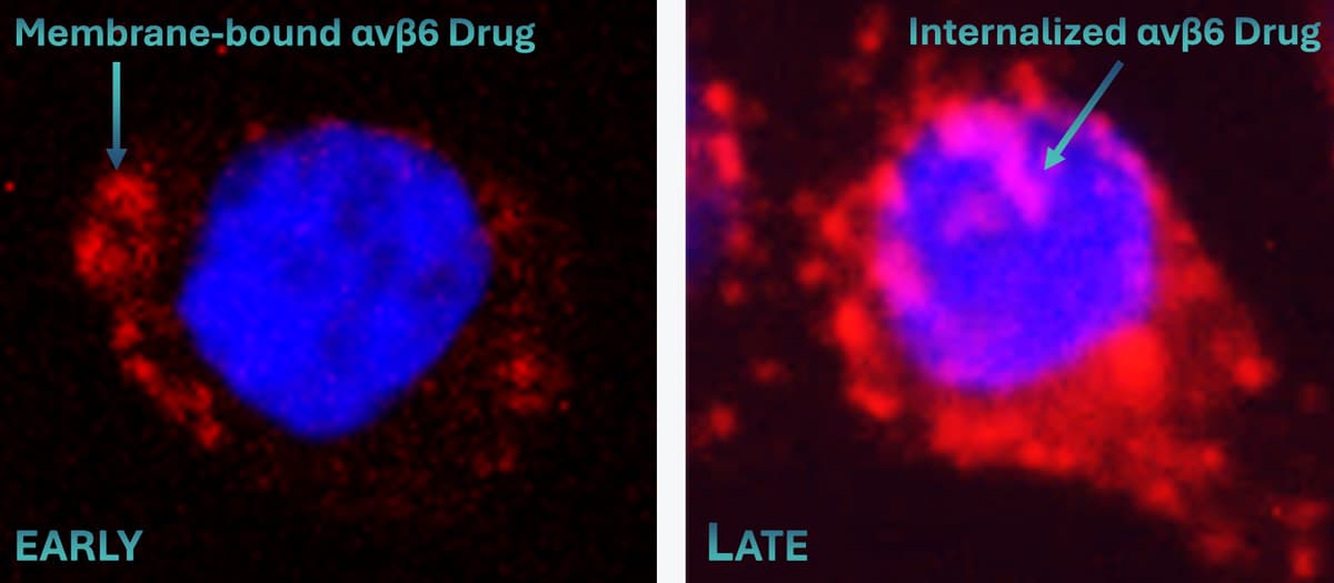
The cell surface protein αvβ6-integrin can be targeted with TRIMT's proprietary cyclopeptide ligands. The protein-ligand complex is rapidly internalized into living cells (see Image), enabling delivery of various payloads to αvβ6-expressing organs or tissues, such as imaging reporters, drugs, or radionuclide warheads against cancer.
αvβ6 - The "Cancer Integrin"
αvβ6-Integrin is expressed by a variety of carcinomas; hence its denomination as "Cancer Integrin".[1] Among them are the most lethal cancer varieties, such as pancreatic ductal adenocarcinoma (PDAC; 88% of cases are αvβ6-positive).[2]
A high prevalence of αvβ6-integrin expression has furthermore been confirmed for other carcinomas, notably non-small-cell lung cancer (NSCLC; 97%), bladder cancer (TCC, 92%), esophageal squamous cell carcinoma (ESCC, 99%), as well as the female cancers endometrial- (EC, 92%), ovarian- (OVCa, 88%), and triple-negative breast carcinoma (TNBC; 89%) (see Image).
αvβ6-Integrin recognizing therapeutics and radioligands have great potential to selectively target and kill the respective tumor cells, while sparing healthy cells. Our goal is to use our proprietary peptides to drive the development of more effective cancer therapies with fewer side effects in order to improve the survival and quality of life of cancer patients.
[1] Quigley NQ, et al., Eur. J. Nucl. Med. Mol. Imaging 2022;49:1136.
[2] Steiger K, et al., Molecular Imaging 2017;16:1536012117709384.
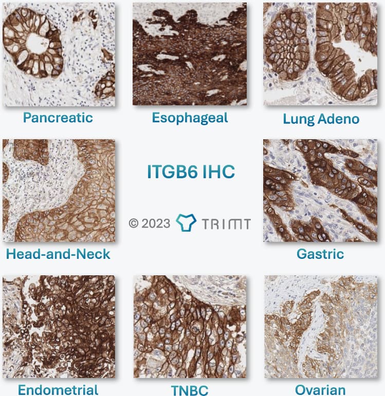
β6-Integrin (ITGB6) Immunohistochemistry (IHC) of tissue sections of various human cancers. In these microscopic images, the blue dots are cell nuclei. Brown color indicates ITGB6 expression. The brown grid-like structures are ITGB6-positive cell membranes. The light-colored areas with smaller nuclei do not contain cancer cells but belong to the microenvironment of the tumor, the stroma.
68Ga-Trivehexin®
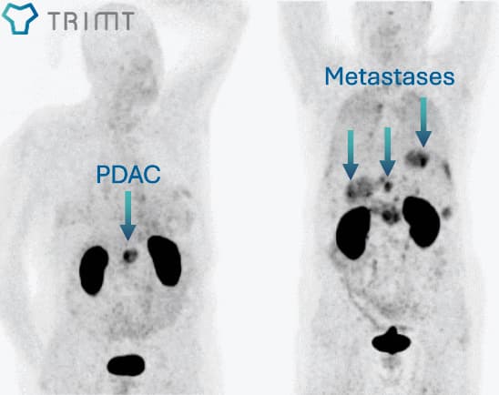
68Ga-Trivehexin® is a radiolabeled bioligand with high affinity for αvβ6-Integrin (a so-called tracer). It contains the short-lived medical isotope Gallium-68 (68Ga) for imaging with positron emission tomography (PET). Since many cancer cells display αvβ6-integrin on their surface, 68Ga-Trivehexin® enables visualization of these cancers by PET, for example, the highly aggressive pancreatic ductal adenocarcinoma (PDAC; see Image).[1,2]
αvβ6-Integrin is expressed by a wide variety of carcinomas and furthermore by fibrotic injuries of the lung, e.g., in the course of idiopathic pulmonary fibrosis (IPF) and COVID-19.[3] 68Ga-Trivehexin® thus also holds great promise for imaging of carcinomas as well as fibrosis and long-covid related pulmonary lesions.
68Ga-Trivehexin® is currently evaluated in clinical trials:
- US Phase 1[4] in healthy volunteers and PDAC
- Single-centre investigator initiated prospective Phase 2[5] for comparison of 68Ga-Trivehexin® and [18F]FDG in PDAC and HNSCC incl. validation of ITGB6 expression by IHC
- in various special national programs for TNBC, PDAC, NHSCC, OVCa, and others.
Beyond its potential for PET imaging of carcinomas and other conditions, 68Ga-Trivehexin® may prove useful as a selection biomarker for therapeutic methods that rely on αvβ6-integrin expression, such as radio-theranostics, targeted drug delivery, or guided surgery.
[1] Quigley NQ, et al., Eur. J. Nucl. Med. Mol. Imaging 2021;48:4107.
[2] Quigley NQ, et al., Eur. J. Nucl. Med. Mol. Imaging 2022;49:1136.
[3] Kimura RH, et al., Am. J. Respir. Crit. Care. Med. 2023 207:1633.
[4] NCT05799274
[5] Thakral P, et al., Cancer Biother Radiopharm. 2023;38:468
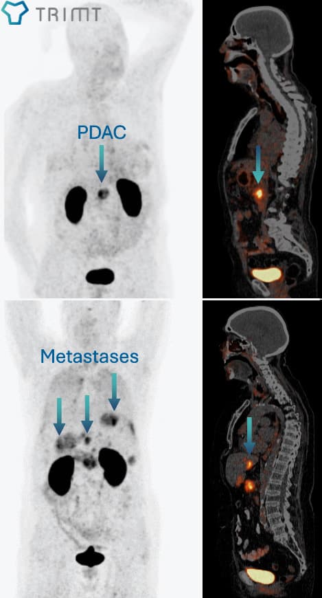
68Ga-Trivehexin® PET/CT images of non-metastatic and metastatic pancreatic ductal adenocarcinoma (PDAC).[1,2]
αvβ6-Integrin Targeted Theranostics
The clinical combination of radiopharmaceuticals for cancer imaging and therapy, frequently referred to as "Radio-Theranostics", involves two steps (see Image):
- Visualization of cancer lesions and selection of patients eligible for therapy by non-invasive imaging techniques such as positron emission tomography (PET).
- Targeted delivery of a radionuclide emitting alpha- or beta-radiation (i.e., energetic particles) to cancer cells, in order to eradicate the cancer lesions by irradiation from the inside.
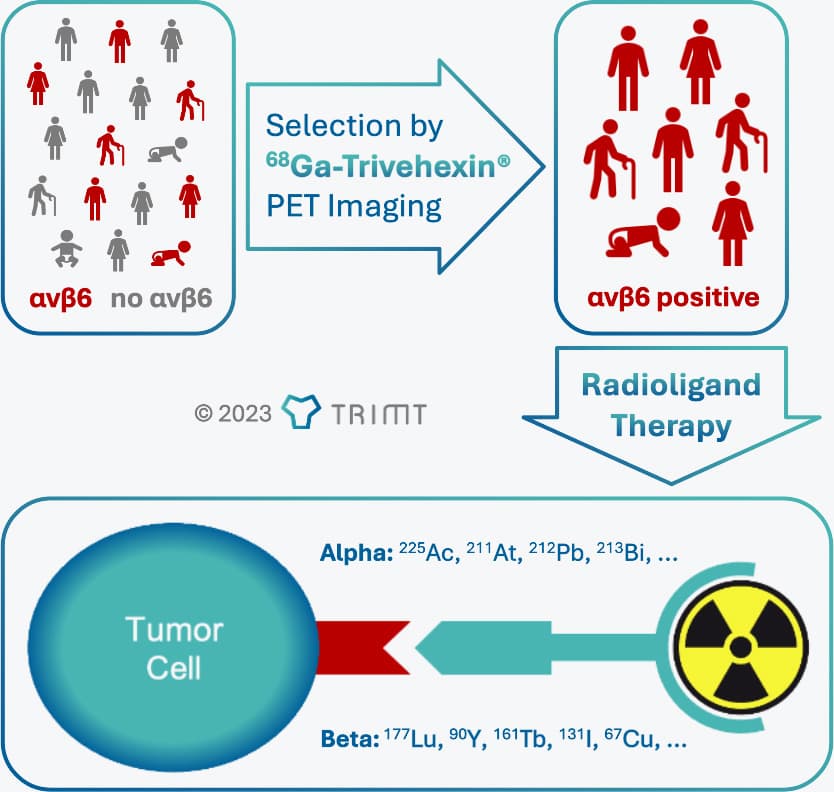
Principle of αvβ6-integrin targeted radio-theranostics, using 68Ga-Trivehexin® as a selection biomarker.
TRIMT's 68Ga-Trivehexin® is a highly specific PET tracer for imaging of αvβ6-Integrin, enabling patient selection for αvβ6-targeted therapies.[1,2] It is thus recommended as selection biomarker for any form of αvβ6-Integrin targeted treatments, including, but not limited to, radioligand therapy.
On the basis of our proprietary αvβ6-integrin selective peptides, TRIMT is actively developing matched therapeutic radiopharmaceuticals to treat eligible cancer patients. Optimized drug candidates are currently prepared for clinical trials.
[1] Quigley NQ, et al., Eur. J. Nucl. Med. Mol. Imaging 2022;49:1136.
[2] Quigley NQ, et al., Eur. J. Nucl. Med. Mol. Imaging 2021;48:4107.
Examples for 68Ga-Trivehexin® PET images of pancreatic ductal adenocarcinoma (PDAC),[1,2] which might be used to identify patients eligible for radioligand therapy.
αvβ6-Integrin Targeted Drug Delivery
The practical possiblity of selecting patients with disease-related, locally elevated αvβ6-integrin expression by using 68Ga-Trivehexin® as a selection biomarker opens an avenue to personalized therapy with highly disease-specific drug formulations (see Image). There is compelling evidence that αvβ6-integrin rapidly internalizes after binding to a ligand, such as a peptide.[1] This mechanism is very robust and works well for peptides that have been equipped with anticancer drugs, such as cytotoxic agents.[2,3]
The respective molecular constructs, often referred to as Peptide Drug Conjugates (PDCs, see Image), have the potential to actively transport the drug component they contain to and into the cancer cells. Such "magic bullets" can achieve higher drug concentrations in tumor tissue than their non-targeted siblings and thus enhance the anticancer effect of a drug. Furthermore, since the higher efficacy of such therapeutics may allow the total amount administered to be reduced, the treatment should be less toxic to αvβ6 integrin-negative healthy cells. PDC-based cancer therapies are therefore potentially more effective and have fewer side effects, with the prospect of improving the survival and quality of life of cancer patients.
World-leading scientists in the field have stated that "it is clear that specific targeting αvβ6 in humans is now a practical possibility and should become a platform for development of improved therapies for the effective treatment of pancreatic and hopefully many other types of cancer."[3] Our goal is to unleash the therapeutic power of PDCs based on our proprietary αvβ6-integrin binding peptides.
[1] Meecham A, et al., Front. Cell Dev. Biol. 2022;10:920303.
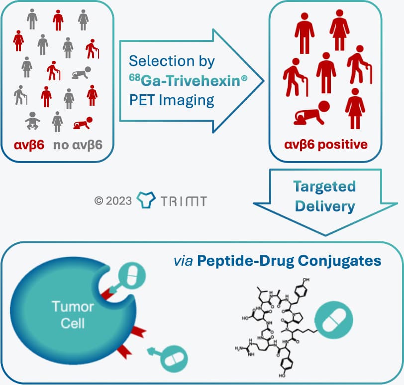
Pharmaceutical intervention enhanced by αvβ6-integrin targeting.
Molecular Imaging & Therapy
TRIMT's proprietary αvβ6-integrin targeting cyclopeptides can be equipped with a wide variety of other functional molecules. These so-called peptide conjugates may comprise fluorescent dyes that can be detected with fluorescence microscopes for cellular imaging. When such a compound is applied during a surgical procedure and surgeons use intraoperative fluorescence camera devices that allow them to better distinguish αvβ6-integrin expressing tumors from surrounding healthy tissue, this is called Fluorescence-Guided Surgery. In case the molecule contains a radioisotope that emits gamma-radiation and the intraoperative tumor detection is done with a handheld gamma probe, the procedure is called Radio-Guided Surgery.
The light-sensitive part of the peptide conjugate can also be a photocatalyst that releases cytotoxic substances (frequently, reactive oxygen species, ROS) when irradiated with light. In practice, such conjugates may be administered like the positron emission tomography (PET) tracer 68Ga-Trivehexin®. One they have reached their target, i.e., the αvβ6-integrin expressing tumor cells, the entire tumor area can be irradiated with high-intensity laser beams to selectively eradicate the tumor while spare the surrounding normal tissue. This concept is widely referred to as Photodynamic Therapy.
Our vision is to advance these emerging technologies for clinical cancer management by developing suitable innovative compounds based on our proprietary αvβ6 integrin-targeting peptides to offer new perspectives to cancer patients.
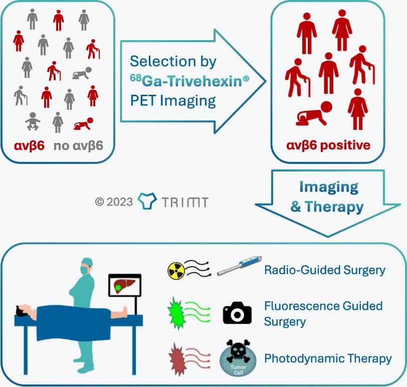
Therapeutic procedures combining αvβ6-integrin targeting and electromagnetic radiation (gamma, optical, IR)
PD-L1 – Immune Checkpoint
PD-L1 is the abbreviation for "Programmed cell Death Ligand 1", a cell surface protein that is recognized by the receptor PD-1 (Programmed Death receptor 1) of certain immune cells. Some cancer cells display PD-L1 in high concentrations on their surface to prevent an attack by the immune system (a so-called immune evasion mechanism).[1] The clinical importance of PD-L1 arises from the fact that immune evasion can be reverted by anti-PD-L1 monoclonal antibodies (mABs) such as durvalumab, atelizumab, or avelumab, or by anti-PD-1 mAbs such as nivolumab or pembrolizumab. The immune system is thus re-enabled to recognize and eradicate the cancer cells. This form of cancer immunotherapy is referred to as checkpoint inhibitor therapy (recognized with the Nobel Prize in Physiology or Medicine, awarded 2018 to the PD-1 discoverer Tasuku Honjo).[2]
Since PD-L1 is only expressed by a fraction of cancers, agents for clinical imaging of PD-L1 are of high interest to select those patients that are likely to benefit from said immunotherapies,[3] or to monitor the progress of ongoing therapies.[4]
TRIMT's portfolio includes tracers for positron emission tomography (PET) imaging of PD-L1, labeled with the short-lived medical isotope Gallium-68 (68Ga). Preclinical imaging confirmed favorable properties for sensitive imaging of PD-L1 expression by PET (see Image).[5]
[1] Ribas A, Wolchok JD. Science 2018;359:1350.
[2] https://en.wikipedia.org/wiki/Tasuku_Honjo
[3] Krutzek F, et al., Pharmaceuticals 2022;15:747.
[4] Nimmagadda S, Cancers 2020;12:3173.
[5] Notni J., et al., unpublished results.
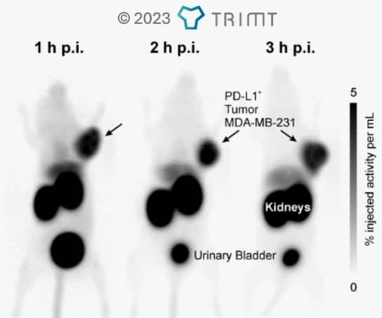
Preclinical PET imaging of PD-L1 expression in the non-transfected PD-L1 positive MDA-MB-213 tumor model with TRIMT's unique 68Ga-labeled PD-L1 PET tracer.[5] Images show a high and persistent signal in the tumor and a generally low background that decreases over time.
Integrin αvβ8 – 68Ga-Triveoctin
αvβ8-Integrin is a potent modulator of the immune system and furthermore – like αvβ6-integrin – an activator of transforming growth factor β (TGFβ). As such, the expression level of αvβ8-Integrin in tissues is connected to a multitude of biological and pathological mechanisms involving TGFβ, including, but not limited to, cancer progression and metastasis.[1]
TRIMT's αvβ8-Integrin targeted positron emission tomography (PET) tracer 68Ga-Triveoctin was developed on the basis of a unique, highly selective octapeptide (see Image).[2] 68Ga-Triveoctin is the first clinically relevant αvβ8-Integrin specific agent of this type.[3] Potential applications include stratification of patients for αvβ8-Integrin-targeted therapies by non-invasive PET imaging.
[1] Takasaka N, et al., JCI Insight 2018;3:e122591.
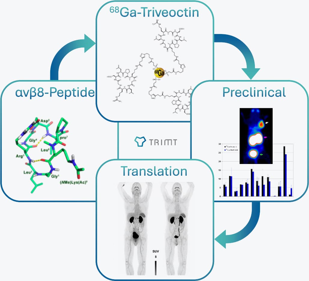
Development course of 68Ga-Triveoctin for PET imaging of αvβ8-Integrin expression.[2,3]